
Oncology
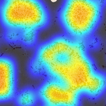
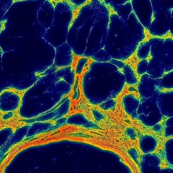
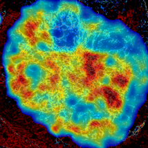
Quantifying fibrosis in the extracellular matrix (ECM) reveals how stromal remodeling drives tumor progression, immune exclusion, and therapy resistance. Objective ECM metrics stratify disease stage and prognosis by capturing collagen density, alignment, and stiffness. Measuring fibrosis guides antifibrotic and immuno-oncology strategies, helping overcome the tumoral fibrotic barrier to immune infiltration and drug delivery.
ONCONEST Quantifies Fibrosis in the context of Oncology
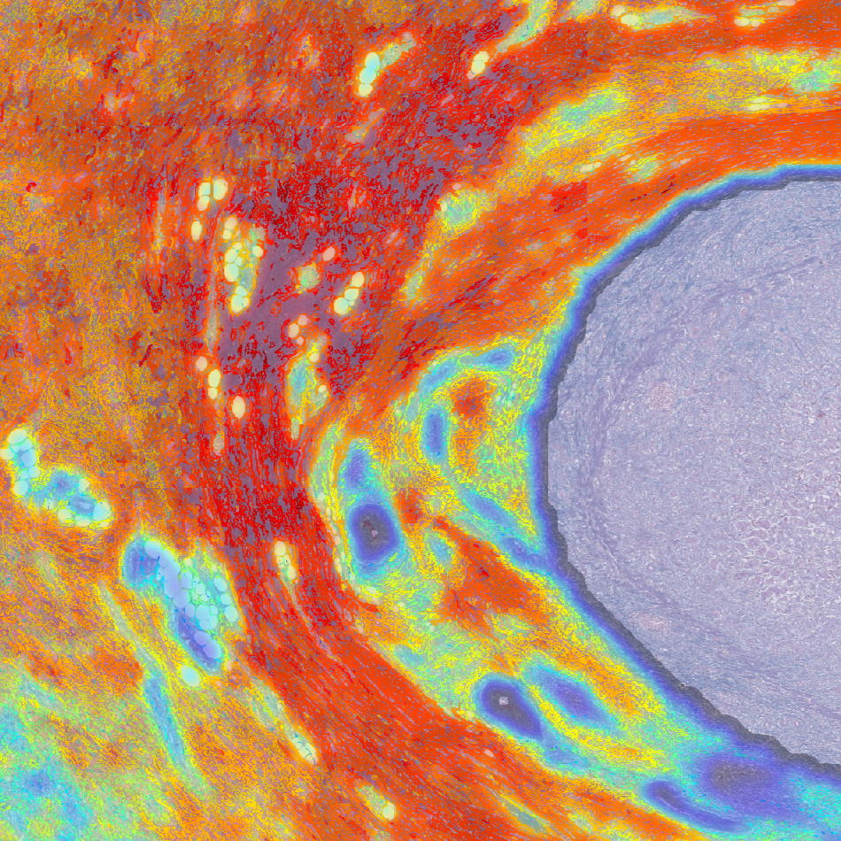
•Intratumoral Fibrotypes
•Peritumoral Fibrotypes
•Tumor burden
•Fibrotic mesh remodeling
•T-Cell percolation through the Fibrotic Capsule
•Biomarkers of severity to improve or complement severity stages
•Biomarkers of recurrence, metastasis, lymph nodes proliferation
FIBROTYPES Distinguish HCC RIKS in PATIENTS WITH NAFLD
Quantitative fibrosis analysis using FibroNest™ identified distinct fibrotypes in cirrhotic NAFLD livers with and without HCC. Automated Ph-FCS scoring differentiated HCC with high accuracy by integrating collagen structure, morphometry, and architecture. This FibroNest approach i poised to enhances early detection, risk stratification, and precision assessment of hepatocarcinogenesis in fibrotic livers.
Automated fibrosis phenotyping of liver tissue from non-tumor lesions of patients with and without hepatocellular carcinoma after liver transplantation for non-alcoholic fatty liver disease. Nakamura Y et al. Hepatol Int. 2022;16(3):555-561. doi:10.1007/s12072-022-10340-9 (weblink)
fibrotypes predict LYMPH NODE METASTASIS and colon cancer outcomes
Quantitative fibrosis phenotyping in advanced colon cancer revealed that digital assessment of collagen structure and organization can accurately predict lymph-node metastasis and recurrence. Using FibroNest™, phenotypic fibrosis composite scores distinguished metastatic and recurrent cases with high sensitivity and specificity, demonstrating fibrosis quantification and fibrotypes as a powerful biomarkers for prognosis and therapeutic stratification.
Intratumoral Fibrotic Features are Associated with Lymph Node Metastasis and Recurrence in Patients with Advanced Colon Cancer. Mine et al. Modern Pathology - Volume 38, Issue 11, November 2025, 100828 - doi.org/10.1016/j.modpat.2025.100828
Fibrosis Patterns as Prognostic OF Cohesive Gastric Carcinoma
FibroNest reveals distinct collagen phenotypes associated with lymph node metastasis and recurrence in advanced colon adenocarcinoma.
A composite fibrosis score integrating fiber morphometrics accurately discriminated metastatic status and post-chemotherapy recurrence demonstrating that objective fibrosis quantification provides a powerful, automated prognostic tool beyond conventional histopathology
Intratumoral Fibrosis Patterns as Prognostic Indicators in Poorly Cohesive Gastric Carcinoma
IN REVIEW
Gastric Cancer, Shigaki et al Yuko Azegawa Ericsson, Valerie Maria E Teves
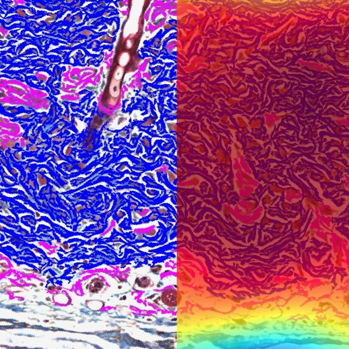
CCN1-Driven Fibrosis Fuels Melanoma Progression
Cancer-associated fibroblast–specific CCN1 expression promotes melanoma progression by enhancing angiogenesis, collagen organization, and metastatic potential. Loss of CCN1 disrupted ECM structure and vascular formation, reducing metastasis. These findings highlight stromal CCN1 and high-resolution fibrotypes as valuable tools for identifying therapeutic targets and predicting resistance to immunotherapy.
Cancer-associated Fibroblast–specific Expression of the Matricellular Protein CCN1 Coordinates Neovascularization and Stroma Deposition in Melanoma Metastasis. Hutchenreuther et Al. Cancer Research Communications (2024) 4 (2): 556–570.