
SSc and Autoimmune interstitial diseases
Autoimmune interstitial diseases such as SSc-ILD, RA-ILD, Sjögren’s-, lupus-, and overlap-associated ILD each feature distinct fibrotic remodeling that can be studied independently. As biopsies are not routine in the clinical management of lung fibrosis (but are in the context of Sjogren’s or SSc), fibrosis burden is evaluate in lung in the context of preclinical studies only. In SSc or Sjogren’s, quantitative histological endpoints provide objective fibrosis endpoints that complement imaging, support non-invasive biomarker development, and enhance disease stratification and therapeutic assessment in both pre-clinical and clinical studies.
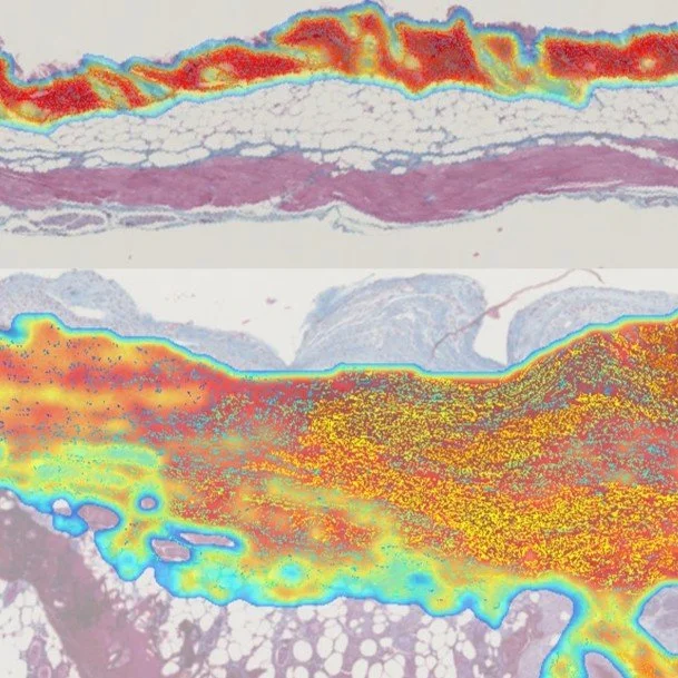
FibroNest Captures Regression Traits in a HOCl SSc Model
Single-fiber digital pathology provides high-sensitivity, objective quantification of fibrotic remodeling in a HOCl mouse model of systemic sclerosis. The AI-derived Phenotypic Fibrosis Composite Score (Ph-FCS) integrates collagen content, structure, and architecture, enabling precise assessment of fibrosis severity and progression. This method outperforms conventional histology, enhancing translational and preclinical fibrosis evaluation.
Single-fiber Digital Pathology Image Analysis accurately quantifies the severity of the fibrosis phenotype in an experimental rodent model of systemic sclerosis.Silvia Speca et Al. Keystone Fibrosis 2023
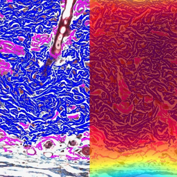
Bridging IPF Fibrotypes FROM RODENT TO HUMAN TISSUES
Single-fiber histological assessment was performed on lung tissues from mice, rats, and patients with idiopathic pulmonary fibrosis (IPF). Quantitative digital analysis revealed shared fibrosis phenotypes—“translational fibrotypes”—spanning species. These conserved morphometric and architectural traits were integrated into a unified translational biomarker, bridging preclinical and clinical fibrosis evaluation.
Comparison of histological phenotype by FibroNest of human IPF and pre-clinical rodent models of lung fibrosis. Francesca Ruscitti et Al, International Colloquium on Lung and Airway Fibrosis, October 12-16 2024:
Fibrosis biomarkers in SSC complement biological findings
Integrating AI-driven digital pathology with biological biomarker co-analysis in systemic sclerosis enables precise quantification of skin fibrosis, vascular remodeling, and immune activation. Linking histologic architecture with circulating and tissue biomarkers is poised to improve disease stratification, prognostic assessment, and treatment-response evaluation.
Cancerassociated Fibroblast–specific Expression of the Matricellular Protein CCN1 Coordinates Neovascularization and Stroma Deposition in Melanoma Metastasis. Hutchenreuther et Al. Cancer Research Communications (2024) 4 (2): 556–570.

Digital Fibrosis Scoring Advances Sjögren’s Diagnosis
In 2023, PharmaNest introduces the first quantitative digital pathology score for fibrosis in Sjögren’s syndrome (SS). By analyzing over 400 collagen traits, FibroNest discriminated SS from controls with exceptional sensitivity and dynamic range. This continuous, image-based biomarker enhances diagnostic precision in a complex Auto Immune fibrotic condition and is poised to supports targeted therapeutic development.
Evaluation of the performance of a novel Digital Pathology score for the evaluation of Fibrosis in patients with Sjögren’s syndrome. Fowler et Al. Keystone Fibrosis 2023